Iron Brain
Behind the comic
What is your research about - in one sentence?
We are developing new ways to use magnetic resonance imaging (MRI) to see how metals such as iron accumulate in the brain, helping to monitor brain health and disease.
What does the comic show?
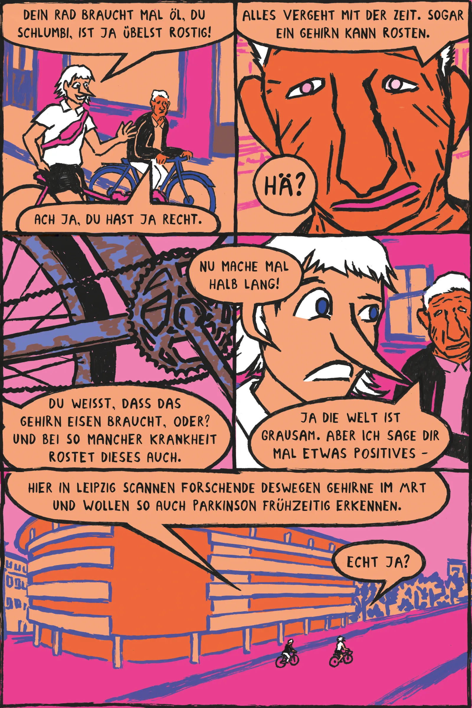
Our comic features a young person and an older person cycling through Leipzig. The older person's rusty bike sparks a conversation about the brain that can also “rust”. At first, the older person offers a pessimistic view of the world, but then he mentions that researchers in Leipzig target this brain rusting. This happens just as they cycle past the Max Planck Institute for Human Cognitive and Brain Sciences.
What findings support this idea?
A healthy brain needs the right amount of iron. Iron plays an essential role in helping the brain cells to communicate to each other, to build the protective coating that surrounds nerve fibers, and to support the brain’s energy supply. Especially in childhood, sufficient iron is important for the proper development and function of the brain. However, as we get older, iron slowly builds up in certain brain regions, and too much iron in cells can trigger harmful chemical reactions, so in a sense, brain cells can "rust". In Parkinson’s disease, for example, the cells that coordinate and fine tune our bodily movement accumulate unusually high levels of iron before they die. The loss of these cells leads to the characteristic movement difficulties associated with the disease. So, both too little iron in childhood and too much iron in old age may be harmful, and it would be helpful for physicians to be able to measure how much iron is in which cells. Because the brain is hidden inside the skull, we cannot simply take a sample to see how much iron is present. Magnetic resonance imaging solves this problem: it creates images of the brain without any surgery and can detect iron indirectly. MRI is sensitive to the magnetic properties of the tissue and as iron is magnetic, it can be measured using MRI.
What are the limits and common misunderstandings?
We are using a powerful MRI scanner as a super microscope to reveal the texture and bio-chemistry of tiny brain regions. However, even the resolution of our most powerful scanners is not high enough to image individual cells in a living person. To this end, we use mathematical modeling to predict measures on the cellular level from the MRI images. We are not using the MRI scanner to treat Parkinson’s or other neurodegenerative diseases, but to develop methods to detect cells at risk early in the disease, so new therapies can be developed. We showed that the measurement works reliably in post mortem tissue but further research is necessary to demonstrate that it works equally well in the living human brain. As part of the international network of researchers “HISTOPARK”, we are currently exploring how well this marker works in vivo.
What questions are still unanswered?
What is the best way to distinguish iron in different cell types and in different forms – the “good” and the “bad” iron? How can we develop useful metrics in the brain that visualize different aspects of the brain’s cellular structure? How can we translate the method from our very powerful research scanners to clinical scanners with a weaker magnetic field and coarser resolution to detect early signs of Parkinson’s disease? How can we prevent important cells from experiencing iron overload while maintaining iron levels necessary for proper brain function? How early and how well can we predict the risk of Parkinson’s disease in an individual using MRI?
How could this shape future medicine?
Iron plays a pivotal role in healthy brain development, normal function and in many neurodegenerative diseases. We develop methods to monitor the brain iron levels across the lifespan without surgery by turning MRI into a quantitative, diagnostic instrument. Iron is a key player in Parkinson’s disease, a slowly progressing neurodegenerative disorder that affects the brain cells many years before the loss of motor control. Our study is expected to enhance our understanding of the early dynamics of Parkinson’s disease and related disorders. MRI can open a window onto the brain’s iron levels, helping both basic scientists and clinicians to evaluate brain health from childhood through old age. By detecting the cell-specific iron overload early, we hope to diagnose and eventually intervene in neurodegenerative diseases before a bike ride becomes a struggle.
What societal and ethical questions does this raise?
Our research advances the understanding of the living brain by using MRI. A great opportunity of MRI is to catch Parkinson’s disease in its silent stage, when protecting brain cells might still be possible. And beyond Parkinson’s, the same technology could help understand aging, depression, or other brain disorders where early changes hide in plain sight. Our project is not just about technology. It connects people, too: patients, families, and researchers across Europe and Canada are interacting and working together. Volunteers take part in gentle sleep tests, give small DNA samples, and return for follow-up scans over several years. Their data — millions of brain pixels, sleep rhythms, and genetic patterns — are combined using computer models that learn to recognize early warning signs.
How do you study this topic?
We use the MRI scanner as a device to measure the physical and biological properties of the brain. We call these methods quantitative MRI in contrast to the conventional MRI in the clinics. Using our advanced MRI methods, we image the brains of a large group, including healthy participants, patients with sleep disorders, and individuals recently diagnosed with Parkinson’s disease. We will utilize newly developed algorithms for image analysis and advanced statistical models to detect small changes in brain structures that regulate sleep and motion. Additionally, we examine donated brains from deceased individuals to precisely map these small regions in the brain. By integrating techniques from anatomy, medical physics, and statistics, we aim to gain new insights into the brain processes of Parkinson’s disease patients.
Where can I learn more about this topic?
More information can be found at our website: https://www.cbs.mpg.de/neurophysics/third-party-funding/histopark
A video explaining how MRI can help us diagnose and treat neurodegenerative diseases can be found here https://www.cbs.mpg.de/departments/neurophysics/gallery and in full length here:
References
Weiskopf, N.; Edwards, L. J.; Helms, G.; Mohammadi, S.; Kirilina, E. Quantitative Magnetic Resonance Imaging of Brain Anatomy and in Vivo Histology. Nat. Rev. Phys. 2021, 3 (8), 570–588. https://doi.org/10.1038/s42254-021-00326-1.
Zecca, L.; Youdim, M. B. H.; Riederer, P.; Connor, J. R.; Crichton, R. R. Iron, Brain Ageing and Neurodegenerative Disorders. Nat. Rev. Neurosci. 2004, 5 (11), 863–873. https://doi.org/10.1038/nrn1537.
Ward, R. J.; Zucca, F. A.; Duyn, J. H.; Crichton, R. R.; Zecca, L. The Role of Iron in Brain Ageing and Neurodegenerative Disorders. Lancet Neurol. 2014, 13 (10), 1045–1060. https://doi.org/10.1016/S1474-4422(14)70117-6.
Brammerloh, M.; Morawski, M.; Friedrich, I.; Reinert, T.; Lange, C.; Pelicon, P.; Vavpetič, P.; Jankuhn, S.; Jäger, C.; Alkemade, A.; Balesar, R.; Pine, K.; Gavriilidis, F.; Trampel, R.; Reimer, E.; Arendt, T.; Weiskopf, N.; Kirilina, E. Measuring the Iron Content of Dopaminergic Neurons in Substantia Nigra with MRI Relaxometry. NeuroImage 2021, 239, 118255. https://doi.org/10.1016/j.neuroimage.2021.118255. https://physics.aps.org/articles/v17/101
Brammerloh, M., Sibgatulin, R., Herrmann, KH., Morawski M., Reinert T., Jäger C., Müller R., Falkenberg G., Brückner D., Pine, K, Deistung, A., Kiselev, V., Reichenbach, J.R., Weiskopf, N. and Kirilina, E. In Situ Magnetometry of Iron in Human Dopaminergic Neurons Using Superresolution MRI and Ion-Beam Microscopy Phys. Rev. X 2024, 14, 021041 https://doi.org/10.1103/PhysRevX.14.021041
Where it's set
About the Project
Science Streets ist ein Wissenschaftskommunikationsprojekt, das Wissenschaft in den Alltag bringt, indem es Leipzigs öffentliche Räume zu Lernorten macht. Für vier Wochen im August 2026 werden Science-Comics auf Werbeflächen (Litfaßsäulen, City-Light-Postern, Infoscreens, im öffentlichen Nahverkehr usw.) gezeigt. Das diesjährige Thema lautet Neurowissenschaften. Zehn Wissenschaftler*innen und zehn Illustrator*innen werden ausgewählt, um gemeinsam Comics rund ums Gehirn zu gestalten – die Wissenschaftler*innen liefern die Inhalte, die Illustrator*innen setzen diese künstlerisch um.
Videos
More Comics